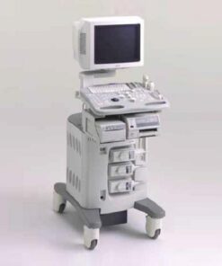
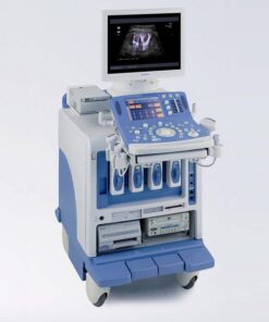
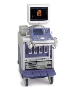
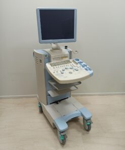
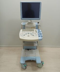
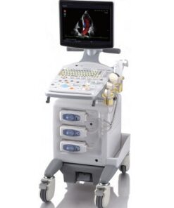
FUJIFILM ARIETTA 50 LE – Ultrasound System
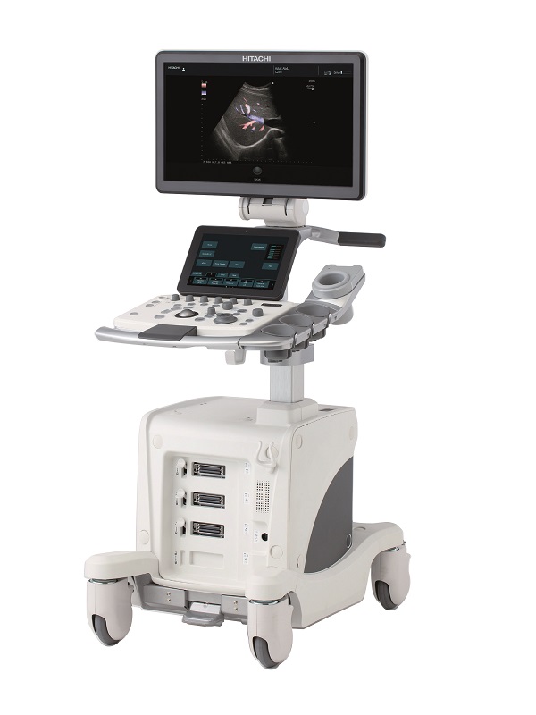
ARIETTA 50 LE Ultrasound is the compact entry model of the ARIETTA series, inviting easy operation from beginners through experts, taking you to the ‘next level’.
Description
FUJIFILM ARIETTA 50 LE – Diagnostic Ultrasound System
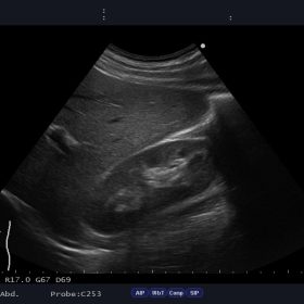
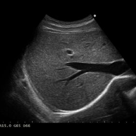
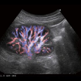
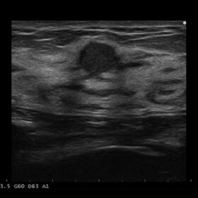
Ultrasound ARIETTA 50 LE – Image Gallery and Videos
Cardio 2D Mode
Cardio Color Mode
Cardio PW, CW and TDI Doppler
Product video
The next level in usability
ARIETTA 50 LE Ultrasound is the compact entry model of the ARIETTA series, inviting easy operation from beginners through experts, taking you to the ‘next level’.
This ultrasound platform combines
- Carefree Workflow,
- Clear Imaging
- Clean Applications
The intuitive workflow of the ultrasound platform allows the operator to focus more on the patient than on the actual operation
- reducing fatigue and musculoskeletal disorders of the examiner
- facilitating examination in various clinical settings by adjusting the platform
ARIETTA 50LE benefits from:
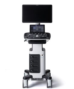
- High contrast 21.5” widescreen LCD monitor to display images with high sensitivity and resolution
- 10.1” touch screen panel, mounted at a comfortable and convenient angle
- Customizable screen layout, selectable by clinical application to allow intuitive operation
- Simplified operating console to speed up your daily routine for a more pleasant operating experience
- User-friendly interface from the home screen, allowing to intuitively input the patient ID, select the examination area and retrieve previously saved image settings
Intelligent Automatic functions for smooth workflow
- Auto Optimizer – to automatically adjust the gain and baseline position and velocity range with just one button
- Mobility that meets autonomy – allowing to scan approx. 1 hour in battery mode for smooth use in emergency care or changing rooms
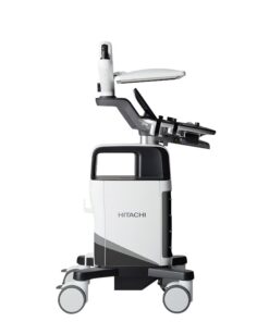
- Power cord hook, handles, and folding mechanism, helping to move the system safely
Clear Imaging for enhanced precision in a wide field of clinical applications
ARIETTA 50 features high-performing image processing technologies, inherited from the ARIETTA series, such as
- High Resolution Color Flow (eFLOW) for detailed depiction of blood flow dynamics and accurate delineation of both, fine and larger blood vessels.
- Silky Image Processing (SIP) to emphasize tissue structures and contrast resolution for clear images
- Compound Imaging for clear visualization of tissue boundaries, high contrast resolution and speckles reduction, allowing a confident observation of lesions
Clean Application featuring intuitive clinical functions, allowing you to concentrate on your patient
- Trapezoidal scanning for wider field of view with linear probes, enhancing visualization of vessels, organs and surrounding tissues
- Free Angler M-mode (FAM) using any cursor orientation to compare wall and valve movement from multiple angles in the same heartbeat
- Auto IMT to automatically measure max and mean values of Intima-Media Thickness (IMT) for reproducible and accurate follow-up of vascular diseases
- Doppler Auto Trace for real-time values of peak velocity and blood vessel resistance (PI, RI)
Additional information
| Weight | 64 kg |
|---|---|
| Dimensions | 77 × 53 × 132 cm |
| Brand | |
| Condition | |
| Μedical specialty | |
| Basic functions | Adaptive Image Processing (AIP), Auto-optimizer, B steer, Compound, M mode, Needle Emphasis, Silky Image Processing (SIP), Tissue Harmonic Imaging (THI), Trapezoidal Scan |
| Colored | |
| Doppler | CW Color Doppler, PW Color Doppler, Real-time Doppler Auto Trace |
| Cardiac functions | ECG module, FAM (Free Angular M-mode), Tissue Doppler Imaging (TDI) |
| Radiology functions | |
| Obstetric functions | |
| Display technology | |
| Image quality | |
| Screen dimensions | |
| Touch Panel | |
| Connectivity | Analog Video Input/Output, DICOM, DVD External, DVI-D, Ethernet, HDMI, USB 2.0 |
| Printing | Ethernet Color Printer, Thermal B/W Printer, USB Color Printer |
| System Portability | |
| Battery | |
| Probe ports | Electronic scanning probes: 3 active, Independent probes: 1 active |
Product Files
Product Files
Compatible Probes
Compatible Probes
Convex
Hitachi C253 Abdominal Convex
Endocavity
Hitachi C41V1 Endocavity Vaginal
Linear
Hitachi L442 Vascular Linear
Hitachi L55 Breast Linear
Hitachi L53K Intraoperative Linear
Phased Aray
Hitachi S11 Phased Array
Independent CW Doppler
Aloka UST-2265-2 Independent CW Doppler

 Ελληνικά