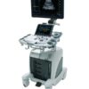
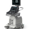
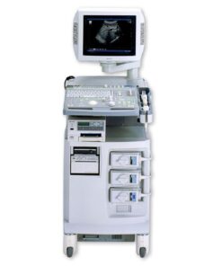
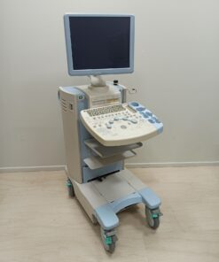
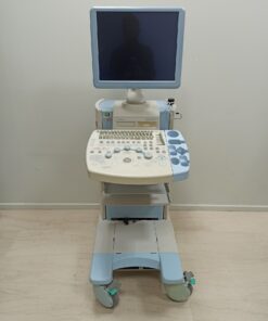
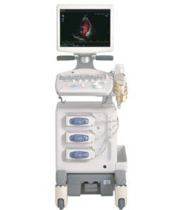
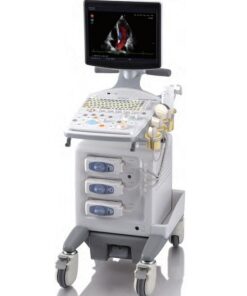
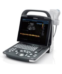
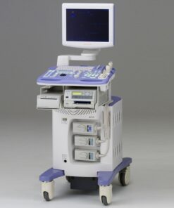
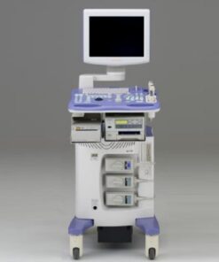
FUJIFILM ARIETTA S60 – Ultrasound System
To provide scanning comfort for all ultrasound users and their patients, the ARIETTA S60 has been designed with ultimate usability.
Description
FUJIFILM ARIETTA S60 – Diagnostic Ultrasound System
Ultrasound ARIETTA S60 – Image Gallery and Videos
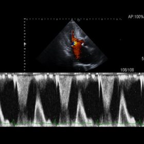
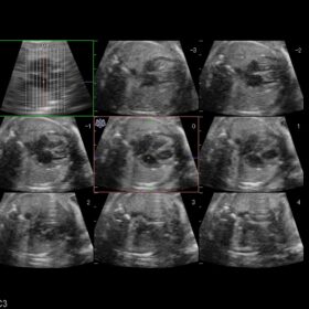
Cardio 2D, Color, PW, CW and TDI mode
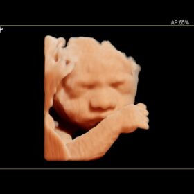
Obstetrics 2D, Color and 4D mode
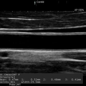
Radiology 2D, Color, Contrast and Elasto mode
Product video
Next Generation Ultrasound Platform
FUJIFILM ARIETTA S60 Ultrasound is equipped with multiple functions dedicated to each diagnostic field, supporting more accurate and efficient diagnoses.
From the extensive range of available transducers, you can be sure to find the one that is appropriate for your examination.
- Less patient-dependent B-mode image variability is experienced; imaging of high quality and sensitivity is available immediately the transducer is applied reducing the need for time-consuming adjustment.
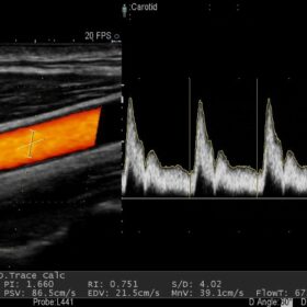
- Blood flow information can be easily acquired. The sharply delineated Doppler waveform is easy to measure.
- Real-time Tissue Elastography (RTE) visualizes relative tissue stiffness, offering complementary information for diagnosis. It has proven clinical value across a wide breadth of applications including breast, thyroid, liver, and prostate.
- Hitachi ARIETTA S60 is equipped with easy-to-use tools for comprehensive cardiac exams: Dynamic Slow-motion Display is a side-by-side presentation of a real-time image and its slow-motion counterpart. Dual Gate Doppler allows observation and measurement of Doppler wave forms from two separate locations during the same heart cycle. These advanced functions can shorten exam times and support effective investigations.
- In obstetrics, the 4D display is a communication tool which can encourage the bond between mother and fetus, and strengthen family ties.
To provide scanning comfort for all ultrasound users and their patients, the ARIETTA S60 has been designed with ultimate usability.
- Hitachi ARIETTA S60 can be moved easily and without strain.
- The monitor can be folded forward to secure safe transport without obstructing your view.
- The panel height can be adjusted to fit both the physique of the examiner and the examination style, offering a comfortable working environment in a natural posture whatever the clinical setting.
- The system can be adjusted from a low position of 70 cm, similar to the height of an office desk, up to 100 cm, an appropriate height for scanning while standing.
Efficient Workflow
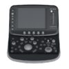
- The operating panel layout of Hitachi ARIETTA S60 is designed with efficient workflow in mind. Free customization of many keys is possible. Probe-dependent assignment of functions to controls offers optimum tailoring for each clinical situation.
- The two-way multi-rotary encoders offer intuitive operation, enabling the management of many functions in one control: working as a joystick for up/down and right/left movement, and as a rotary encoder to adjust other parameters.
- The Auto Optimizer feature corrects many parameters with one key-stroke, instantaneously forming uniform, diagnostic-quality images.
- The Auto Trace function of Doppler waveforms supports exam efficiency significantly reducing the number of required key strokes.
Additional information
Product Files
Compatible Probes
Compatible Probes
Convex
Hitachi C35 Abdominal Convex
Hitachi C251 Abdominal Convex
Hitachi C22P Biopsy Convex
Hitachi C22T Intraoperative Convex
Hitachi C22K Intraoperative Convex
Hitachi C22I Intraoperative Convex
Hitachi C25P Biopsy Convex
Hitachi C41 Convex Pediatric
Hitachi C42 Convex Pediatric
Hitachi C42K Intraoperative Convex
Hitachi C42T Intraoperative Convex
Endocavity
Hitachi C41V1 Endocavity Vaginal
Hitachi C41B Endocavity Vaginal/Rectal
Hitachi C41RP Endocavity Rectal
Linear
Hitachi L34 Vascular Linear
Hitachi L441 Vascular Linear
Hitachi L44 Vascular Linear
Hitachi L55 Breast Linear
Hitachi L64 Vascular Linear
Hitachi L43K Intraoperative Robotic
Hitachi L44K Intraoperative Linear
Hitachi L53K Intraoperative Linear
Hitachi L44LA Intraoperative Linear
Hitachi L44LA1 Intraoperative Linear
Hitachi L51K Intraoperative Robotic
Hitachi L46K Intraoperative Linear
Hitachi L53K Intraoperative Linear
Phased Aray
Hitachi S211 Phased Array
Hitachi S31 Pediatric Phased Array
Hitachi S3ESL1 TEE Cardio
Hitachi S31KP Intraoperative Burr-Hole
RT-3D (4D)
Hitachi VC34 Convex 3D/4D
Hitachi VC35 Convex 3D/4D
Hitachi VL54 Linear 3D/4D
Hitachi VC41V Vaginal 3D/4D
Bi-Plane
Hitachi CC41R Endocavity Bi-Plane
Hitachi CC41R1 Endocavity Bi-Plane
Hitachi CL4416R Endocavity Bi-Plane
Hitachi C41L47RP Endocavity Bi-Plane
Independent CW Doppler
Aloka UST-2265-2 Independent CW Doppler
Aloka UST-2266-5 Independent CW Doppler
Radial
Hitachi R41R Endocavity Electronic Radial
Hitachi R41RL Endocavity Electronic Radial

 Ελληνικά